You may visit the dentist feeling perfectly comfortable and still be told that dental X-rays would be helpful. That is because a visual examination can reveal a great deal about your mouth, but it cannot show everything happening inside a tooth, between teeth, or beneath the gums.
Dental X-rays, also called dental radiographs, provide images that help dentists diagnose and monitor oral conditions that may not yet be visible or causing symptoms.
According to the American Dental Association’s patient guide to dental X-rays, X-rays can reveal conditions such as new cavities and impacted teeth that may not be apparent during a routine clinical examination. The ADA also emphasizes that X-rays should be recommended according to each patient’s individual diagnostic needs rather than automatically taken at every dental visit. MouthHealthy
For patients considering dental X-rays in Anaheim, understanding why an image is being recommended can make this routine diagnostic tool easier to understand.
What Are Dental X-Rays?
Dental X-rays create images of teeth and surrounding structures that cannot be fully evaluated simply by looking inside the mouth.
Different types of dental X-rays provide different views.
Depending on the image being taken, a dentist may be able to evaluate:
- Areas between teeth
- Tooth roots
- Supporting bone
- Developing or unerupted teeth
- Wisdom teeth
- Existing restorations
- Areas of suspected decay
- Changes around the roots of teeth
X-rays supplement the dental examination. They do not replace it.
The dentist combines the images with your symptoms, dental history, clinical examination, and other findings when making a diagnosis.
Why Can’t a Dentist See Every Cavity During an Exam?
Some cavities occur on tooth surfaces that are easy to inspect directly.
Others develop between neighboring teeth, where visual examination may provide limited information.
Dental radiographs can help reveal changes in these areas before the cavity becomes large enough to be obvious from the outside.
This can be particularly useful because tooth decay does not always cause pain during its earlier stages.
Finding decay does not automatically mean extensive treatment will be required. The size, location, and activity of the lesion all matter when determining the appropriate next step.
What Can Dental X-Rays Detect?
Dental X-rays may provide useful information about several oral health conditions.
The ADA notes that radiographs can reveal dental disease or damage that is not visible during a standard examination. MouthHealthy
Depending on the type of X-ray, dentists may use the images to investigate:
- Tooth decay between teeth
- Problems beneath existing restorations
- Impacted teeth
- Tooth development
- Bone surrounding the teeth
- Certain infections around tooth roots
- Tooth and root position
- Some forms of dental trauma
An X-ray by itself does not always establish the complete diagnosis. It is interpreted alongside the clinical examination.
Do You Need Dental X-Rays at Every Checkup?
No.
Current ADA guidance does not support automatically taking the same X-rays at every dental visit.
The ADA states that the need and frequency of dental X-rays depend on factors including:
- Current oral health
- Age
- Risk of dental disease
- Signs or symptoms
- Dental history
In 2024, an ADA expert panel recommended that radiographs be obtained only when the dentist believes they will provide necessary diagnostic information. MouthHealthy
This means two patients attending routine examinations may appropriately have different X-ray schedules.
Why Might a New Patient Need X-Rays?
When you visit a new dental office, the dentist needs to understand your current oral health.
Existing X-rays from a previous dentist may sometimes provide the information needed.
The ADA recommends asking a previous dental office to forward usable X-rays to the new dentist when appropriate. This may help avoid unnecessary duplication. MouthHealthy
However, additional images may still be recommended if existing X-rays are outdated or do not show the areas required for the current examination.
What Are Bitewing X-Rays?
Bitewing X-rays show portions of the upper and lower teeth in the same image.
They are particularly useful for evaluating areas between back teeth, where cavities may be difficult to see directly.
Bitewings can also provide information about the supporting bone around teeth.
The dentist determines whether these images are needed based on the patient’s individual risk and dental history rather than simply taking them according to a universal timetable.
What Are Periapical X-Rays?
A periapical X-ray generally shows an entire tooth from its crown to the end of its root, along with some of the surrounding bone.
This type of image can be useful when a dentist needs more information about a particular tooth.
For example, it may be considered when investigating:
- Persistent tooth pain
- Dental trauma
- A suspected infection
- Root anatomy
- A tooth requiring root canal treatment
The image allows the dentist to evaluate structures below the gumline that cannot be seen during the visual examination.
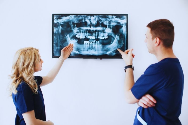
What Is a Panoramic Dental X-Ray?
A panoramic X-ray provides a broad view of the jaws, teeth, and surrounding structures in one image.
Instead of placing a small sensor beside individual teeth, the imaging equipment moves around the patient’s head.
Panoramic images may be useful when evaluating broader anatomical relationships, including the position of developing or impacted teeth.
For example, a panoramic image may be recommended when assessing wisdom teeth and their position within the jaws.
Why Are X-Rays Used Before Some Dental Treatments?
Certain procedures require information about structures that cannot be evaluated adequately from the surface.
For example, X-rays may contribute to planning for:
- Root canal treatment
- Wisdom tooth removal
- Dental implants
- Certain crowns or restorations
- Orthodontic treatment
- Evaluation after dental trauma
The type and number of images should correspond to the information needed for that particular treatment.
More imaging is not automatically better. The objective is to obtain sufficient diagnostic information using an appropriate imaging approach.
Are Dental X-Rays Safe?
Dental X-rays use ionizing radiation, so appropriate use is important.
However, modern dental imaging uses relatively low radiation doses.
The ADA states that the risk of potentially harmful effects from dental radiographs is very small and that advances in dental imaging have substantially reduced radiation exposure. MouthHealthy
Dentists also follow the principle that X-rays should be taken when the expected diagnostic benefit justifies the exposure.
This is another reason individualized imaging is preferred over automatically taking X-rays according to a fixed schedule.
What About Dental X-Rays During Pregnancy?
If you are pregnant or think you may be pregnant, tell your dental team.
Necessary dental care should be discussed with your dentist based on your individual circumstances.
Current ADA patient guidance notes that modern dental X-ray techniques produce low radiation exposure and that expert recommendations have changed regarding routine abdominal and thyroid shielding. MouthHealthy
The dentist can explain why a particular image is being recommended and whether it is necessary at that time.
Why Might the Dental Office Not Use a Lead Apron?
Patients who remember dental visits from previous decades may notice changes in X-ray procedures.
Historically, lead aprons and thyroid collars were commonly used during dental imaging.
The ADA’s current guidance states that abdominal and thyroid shielding is no longer considered necessary for dental X-rays for patients of any age or health status, including pregnancy. One reason is that improperly positioned shielding can interfere with the X-ray beam and require an image to be retaken. MouthHealthy
Some dental practices may still use shielding because of local regulations or other practice considerations.
Can Dental X-Rays Show Gum Disease?
X-rays can provide useful information about the bone supporting the teeth.
This can be important when evaluating periodontal disease because more advanced disease can affect supporting bone.
However, an X-ray is only part of a periodontal evaluation.
The dentist or dental hygienist may also assess:
- Gum inflammation
- Bleeding
- Gum recession
- Periodontal pocket depths
- Plaque and tartar accumulation
- Tooth mobility
Combining clinical findings with appropriate imaging provides a more complete picture of periodontal health.
Can an X-Ray Tell Whether You Need a Root Canal?
Not by itself.
Dental X-rays can provide important information about a painful or damaged tooth, including its roots and surrounding bone.
However, determining whether root canal treatment is necessary can also involve:
- Your symptoms
- Clinical examination
- Temperature testing
- Bite testing
- Other diagnostic procedures
A tooth can sometimes have significant pulp inflammation before obvious changes appear around the root on an X-ray.
Diagnosis therefore depends on the complete clinical picture.
Why Are X-Rays Useful for Wisdom Teeth?
Wisdom teeth can remain partly or completely beneath the gums and bone.
An X-ray can show their position even when they have not erupted into the mouth.
This information may help the dentist evaluate:
- Whether the wisdom tooth is impacted
- Its direction of eruption
- Its relationship to neighboring teeth
- Root development
- Surrounding structures
Not every wisdom tooth automatically needs removal. Imaging helps the dentist evaluate the individual situation and determine whether monitoring or treatment is appropriate.
Can You Request X-Rays From Your Previous Dentist?
Yes.
If you change dentists, ask whether your previous dental office can transfer recent diagnostic images.
The ADA specifically recommends having previous X-rays forwarded when appropriate so a new dentist can determine whether those images provide sufficient current information. MouthHealthy
Your new dentist may still recommend additional images if the existing ones do not answer a current diagnostic question.
Bringing your dental history with you can help the new practice understand changes over time.
What Should You Ask Before Having Dental X-Rays?
Patients should feel comfortable asking why an image is being recommended.
Useful questions include:
- What does this X-ray help you evaluate?
- Are my previous X-rays still useful?
- What type of X-ray are you taking?
- Is the image related to a specific symptom or treatment?
- How will the result affect my treatment plan?
A dentist should be able to explain how the recommended imaging relates to your oral health.
Dental Exams and X-Rays in Anaheim, CA
Dental Aesthetics Professionals provides preventive and general dental care for patients in Anaheim. Dental examinations may include appropriate diagnostic imaging when it is useful for evaluating areas that cannot be fully assessed visually.
If it has been some time since your last dental examination, or you have a tooth that is painful, sensitive, or otherwise concerning, an evaluation can determine whether dental X-rays would provide useful diagnostic information. Having an examination does not mean X-rays or treatment will automatically be necessary; recommendations can be based on your individual dental history and current findings.
Learn more about general dental care at Dental Aesthetics Professionals or call 714-772-3538 to schedule an appointment.